


| Quantity: | |
|---|---|


SF-SWAF10MM-A2
SINCEREFIRST
| This is a mini 5MP USB side-view endoscopic LED camera module, centered on "side-view angle to break through blind spots + high-definition imaging", and suitable for precise detection of target sides/inner walls in medical, industrial, and other fields. The module is equipped with a 1/2.7-inch 5MP CMOS image sensor, which can stably output high-definition images with 5MP (2592×1944) and 1080P (1920×1080) resolutions, and supports a maximum frame rate of 30FPS, providing high-fidelity visual support for side-view observation. The module adopts a "lightweight + anti-complex environment" solution: its cylindrical body with a diameter of 10mm can be easily embedded in narrow spaces. It is equipped with a lens with a fixed 73° side-view field of view (FOV), using a 4P+RI multi-layer coated lens assembly. Combined with auto-focus, it can clearly capture the side/inner wall features of the target; 6 built-in high-brightness LED fill lights of 0402 specification ensure clear details can still be captured in dim environments. In terms of interface, the module uses a standard USB 2.0 Type-A interface and supports the UVC 1.1 protocol, realizing "plug-and-play"—no driver installation is required to be compatible with Windows, Mac OS, Linux, and other systems. After connecting to computers or industrial tablets, it can achieve high-speed data transmission of 480Mbps with video latency of less than 30ms; at the same time, it has passed CE, FCC, RoHS, and ISO certifications, meeting the compliance requirements of medical clinical practice and industrial quality inspection. | 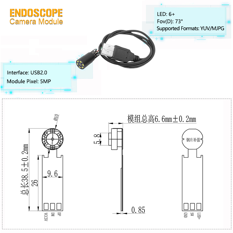 |
Side-View Angle Advantage: The unique side-view lens design effectively avoids blind spots in the front, and can directly observe areas that are difficult for conventional front-view lenses to capture clearly, greatly improving the coverage and accuracy of inspections.
Miniaturized Design: The probe part adopts an ultra-fine outer diameter design of 10mm, with excellent rigidity. It can easily penetrate into narrow pipelines, internal gaps of machinery, and other confined spaces to achieve non-destructive testing.
High-Definition Imaging: The high-definition imaging system built on a 5-megapixel CMOS sensor can output static images with a resolution of 2592×1944. Combined with image processing technology, it can clearly present subtle defects, cracks, or contaminants on the surface of objects.
Plug-and-Play Convenience: Adopting a universal USB interface protocol, it can be recognized by the system without complex driver installation after connection, significantly lowering the threshold for integration and operation. Its compatibility covers mainstream platforms such as Windows, Android, Linux, and Mac OS, facilitating rapid deployment on various devices.
Product Name | 5MP Endoscope Camera Module |
Pixel | 5MP |
Working Current | 140mA-150mA |
Output Interface | USB2.0 |
Image Sensor | CMOS |
Features | Mini Endoscope Camera |
Lens | FOV 73° 4P+RI |
Diameter | 10mm |
Led | 6PCS |
Warranty | 10 Years |
Biliary Tract Probe: As a side-view probe integrated into choledochoscopes, it can observe the location of stone impaction on the bile duct sidewall and the side edge condition of bile duct stenosis after entering through the biliary tract, avoiding missing sidewall lesions due to front-view angle.
Auxiliary Arthroscope: Adapts to side-view auxiliary arthroscopes for minimally invasive surgeries of knee and shoulder joints. It can observe the cartilage wear on the side of joint gaps and the state of ligament attachment points, supplementing the observation blind spots of the main scope and improving surgical accuracy.
Sinuscope: Can penetrate into sinus cavities to observe whether there are polyps, inflammation, or fluid accumulation on the sinus wall mucosa. It solves the problem that front-view lenses are difficult to clearly see the side edges of sinus cavities, assisting otolaryngologists in formulating surgical plans.
A1:It has a fixed 73° side-view field of view (FOV), specifically designed for clear observation of target sides and inner walls.
A2: No. It supports plug-and-play via the UVC 1.1 protocol, so no additional drivers are required.
A3: Yes. It is equipped with 6 high-brightness 0402-spec LED fill lights to ensure clear imaging in low-light conditions.
A4: Yes. It integrates auto-focus technology, enabling precise capture of subtle features on target sides/inner walls.
A5: It works with Windows, Mac OS, Linux, and Android—covering all mainstream platforms for flexible deployment.
A6: Yes. It meets CE, FCC, RoHS, and ISO standards, satisfying compliance requirements for both medical clinical use and industrial quality inspection.


