


| Quantity: | |
|---|---|


SF-U4530-SL-V3
SINCEREFIRST
This 0.3MP USB sensor BF2013 high-definition waterproof endoscopic camera module with LED is specially designed for medical scenarios, providing reliable support for clinical diagnosis and operations with its core parameter advantages. Equipped with the BF2013 CMOS image sensor, it can output clear color images with 0.3MP pixels. Combined with VGA resolution and a maximum frame rate of 30FPS, it can present stable and smooth dynamic images inside cavities in real-time, meeting the needs of detailed observation. The 1/9-inch sensor size ensures imaging quality while helping the module achieve a compact design. The 45° field of view accurately covers the inspection area, avoiding missed inspections due to a too narrow field of view and preventing judgment errors caused by wide-angle distortion. The 15mm focal length combined with manual focusing function can flexibly adapt to the observation of cavities at different depths, ensuring that the details of key lesions are clearly presented. The module adopts an integrated design, and the 4.5mm slender lens diameter can easily enter narrow cavities, reducing patient discomfort. The 6 LED beads integrated in the lens provide sufficient illumination, solving the problem of insufficient light inside the cavity. The bare module produced by SMT process and AA process has both structural stability and assembly flexibility, and can be conveniently adapted to various endoscopic equipment, providing a cost-effective imaging solution for minimally invasive diagnosis and treatment. | 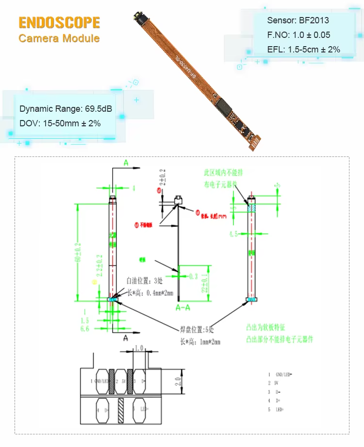 |
Clear and Stable Imaging: 0.3MP pixels combined with VGA resolution and 30FPS frame rate, presenting real-time dynamic images inside the cavity with distinguishable details.
Accurate and Flexible Observation: 45° field of view covers the inspection area; 15mm focal length + manual focusing adapts to the observation of cavities at different depths.
Better Minimally Invasive Experience: Integrated design + 4.5mm slender lens, easily entering narrow cavities and reducing patient discomfort.
Sufficient and Reliable Illumination: Integrated with 6 LED beads to solve the problem of insufficient light inside the cavity.
Strong Adaptability: Produced by SMT process and AA process, with stable structure; the bare module can be flexibly adapted to various endoscopic equipment.
1. Digestive Tract Examination: Adapted to gastroscopes and colonoscopes for observing lesions on the inner walls of the esophagus, stomach, and intestines.
2. Otorhinolaryngology Diagnosis and Treatment: Used in laryngoscopes, otoscopes, etc., for examining inflammation or foreign bodies in narrow cavities such as the throat and ear canal.
3. Minimally Invasive Surgery Assistance: Providing real-time imaging support inside the cavity during minimally invasive surgeries such as laparoscopy and arthroscopy.
4. Gynecological Examination: Adapted to colposcopes for observing subtle lesions in areas such as the cervix.
5. Oral Diagnosis and Treatment: Assisting oral endoscopes in examining the condition of hidden areas such as gums and the adjacent surfaces of teeth.
Product Name | USB Endoscope Camera Module |
Features | BF2013 Endoscope Camera |
Pixel | 0.3MP |
Sensor | BF2013 |
Sensor Size | 1/9 Inch |
Focus Range | 15-100mm |
Focal Length | 3.75mm |
Trademark | SINCERE FIRST |
Fov | 45° |
Diameter | 4.5mm |
Large depth of field (such as 5mm to 50mm) can clearly show near and far objects at the same time, reduce the need for frequent focusing, suitable for endoscopic surgery; Shallow depth of field is used to highlight specific plane details, such as skin inspection.
The medical-grade module uses IP68 waterproof seal, the housing material is resistant to high temperature and pressure (such as epoxy resin), and can withstand 134°C steam sterilization. Industrial modules may require only a splash proof design (IP54).
The medical module emphasizes biocompatibility, sterilization and high image quality, and needs to comply with FDA/CE certification; Industrial modules focus on durability (such as corrosion protection), wide temperature operation (-20°C to 70°C), and special light sources (such as UV).
The cable length is usually 10-30 meters, including Kevlar tensile layer and waterproof connector, which supports real-time transmission while resisting internal friction and chemical corrosion of the pipeline.
Common requirements include special dimensions (e.g., 1mm diameter), multispectral imaging (e.g., fluorescence navigation), and interface protocol adaptation (e.g., interfacing with DICOM systems).
Images of the left and right eyes are captured through a dual lens module or a splitter prism, and synchronously output to a 3D display at ≥1080p/60fps to ensure smooth operation, suitable for minimally invasive surgery.


