


| Quantity: | |
|---|---|


SF-C1019USB-D3.9-PRO-95D V1.2 OV9734
SINCEREFIRST
| This high-performance medical endoscope camera module is built around the OmniVision OV9734 CMOS color image sensor, integrating advanced imaging technology and robust structural design to meet the rigorous requirements of medical diagnosis and treatment scenarios. It features a 1MP pixel resolution, supports 720 resolution output, and delivers a maximum frame rate of 30FPS, ensuring smooth and clear real-time image transmission—critical for capturing dynamic tissue movements during procedures. The sensor, with a size of 1/9 inch and pixel dimensions of 1.4μm×1.4μm, is equipped with OmniVision’s exclusive PureCel® technology. This technology optimizes pixel-level light-sensing efficiency, significantly enhancing light collection capabilities to improve imaging quality in low-light environments—addressing the challenge of dim visibility in deep body cavities. The module offers a precise field of view (FOV) of D95°×H85°×V43.5°, enabling comprehensive coverage of inspection areas while maintaining image clarity without edge distortion (distortion rate <10%). Structurally, it adopts a Separated design that allows flexible integration with different medical devices, simplifying assembly and maintenance for healthcare facilities. The module’s lens is fitted with a Steel Shell, protecting the internal optical components from mechanical damage and ensuring durability during repeated use and disinfection. With an ultra-compact diameter of 3.9mm, it can easily access narrow anatomical spaces while minimizing patient discomfort. It also meets IP67-level dust and water resistance, making it suitable for use in humid clinical environments and compatible with common medical disinfection processes. For connectivity and compatibility, the module uses a USB 2.0 interface and supports the UVC protocol, enabling plug-and-play functionality with mainstream operating systems (Windows, macOS, Linux) without the need for additional drivers—greatly reducing deployment time in medical settings. Manufactured via SMT and AA (Active Alignment) processes, it ensures high optical precision and consistent performance across batches. Additionally, it has passed international certifications including CE, FCC, RoHS, and Reach, verifying its compliance with global safety, electromagnetic compatibility, and environmental protection standards for medical devices. | 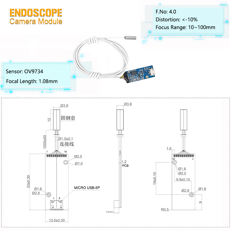 |
Superior Low-Light Imaging Performance: Leveraging the OV9734 sensor’s PureCel® technology and 6 integrated high-brightness LED fill lights, the module effectively reduces noise generation in low-light environments. Even under weak lighting conditions, it captures clean, clear images, ensuring clinicians can clearly observe subtle lesions or tissue details.
Accurate Color Reproduction: The sensor’s optimized color processing algorithm, combined with the high-quality optical lens, accurately reproduces the true colors of human tissues. This intuitive color feedback helps clinicians make more accurate judgments about tissue health status, supporting precise diagnosis and treatment planning.
High-Speed Response & Dynamic Capture: With a maximum frame rate of 30FPS and fast signal transmission via USB 2.0, the module enables high-speed continuous shooting and rapid response to dynamic scenes. It can reliably capture transient tissue movements, avoiding missed critical visual information.
Flexible Separated Design: The Separated structure allows independent installation and maintenance of the module’s imaging unit and control unit, adapting to the layout requirements of different medical devices and improving deployment flexibility.
Robust Protection & Durability: The 3.9mm-diameter lens with a Steel Shell enhances structural strength, while IP67 dust and water resistance ensures the module remains functional after exposure to moisture, dust, or routine medical disinfection. This extends the module’s service life and reduces replacement costs.
High Precision & Compliance Assurance: The SMT + AA manufacturing process guarantees precise alignment between the sensor and lens, minimizing optical deviations and ensuring stable image quality. Compliance with international certifications (CE, FCC, etc.) also eliminates safety and regulatory risks for medical institutions during use.
Wide Compatibility: Support for the UVC protocol and USB 2.0 interface enables seamless connection to various medical workstations, laptops, or display devices. This plug-and-play capability eliminates the need for specialized software development, reducing the technical threshold for clinical application.
Minimally Invasive Surgery: Compatible with laparoscopic and arthroscopic devices—its 3.9mm compact size fits through small surgical incisions, and the high-definition image transmission helps surgeons observe delicate tissue structures for precise operation.
Otolaryngology (ENT) Examination: Ideal for otoscopes and nasoscopes. The ultra-short focal length and narrow 3.9mm diameter allow easy access to narrow ear canals and nasal cavities, reducing patient discomfort while capturing clear images of minor lesions.
Veterinary Diagnosis & Treatment: Used with pet digestive tract endoscopes and respiratory endoscopes to examine the gastrointestinal tract or respiratory tract of companion animals, helping veterinarians identify inflammation, foreign bodies, or ulcers.
Scientific Research Observation: Integrated into microbial sample high-definition recording devices—its low noise and high resolution support clear imaging and data analysis of micro-biological samples, meeting the needs of life science research.
Specialized Medical Auxiliary Detection: Adaptable to small-size gynecological endoscopes or urological endoscopes. The IP67 protection and corrosion-resistant Steel Shell ensure reliability in humid clinical environments, while the PureCel® technology ensures clear imaging even in low-light areas of the body.
Product Name | OV9734 Sensor USB Endoscope Camera Module |
Focal Length | 1.08mm |
DSP | USB |
Sensor | OV9734 |
Focusing Range | 10-30mm |
Diameter | 3.9mm |
F Number | 4.0 |
LED: | 6pcs |
Resolution | 30fps |
FOV(D) | 95° |
1.Why does otorhinolaryngology need ultra-fine diameter modules?
When the space of the ear canal/nasal cavity is narrow, a module with a diameter of ≤3mm can reduce the patient's discomfort. At the same time, it is necessary to maintain high resolution (such as ≥300 line pairs /mm) to identify minor lesions.
2.What could be the possible reasons for noise in an image?
It might be due to sensor overheating, signal transmission interference, or excessively high ISO in low light conditions. It is necessary to check the heat dissipation design, cable shielding, or enable the noise reduction mode.
3.Why does the image show color cast?
It may be due to uncalibrated white balance, mismatched color temperature of the light source (such as mixed use of LED and sunlight), or aging of the sensor. Software correction or replacement of the filter is required.
Our company has several advantages that set us apart from our competitors and make us the first choice among our clients. Here are some of our key advantages:
1. Customer Service: Our team of professionals is loyal and proactive towards customers, responding promptly and effectively solving problems.
2. Timely Delivery: We understand the importance of delivering products on time. Our efficient logistics team ensures that we get our products to our customers on time.
3. Innovation: We continually strive to innovate and improve our products and services. We invest in research and development to stay at the forefront of our industry.


