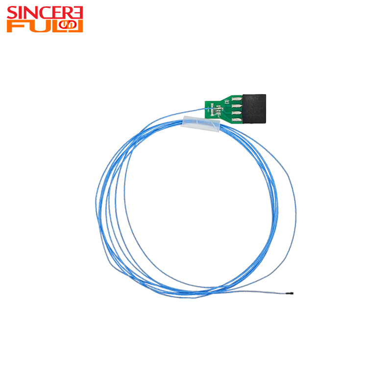
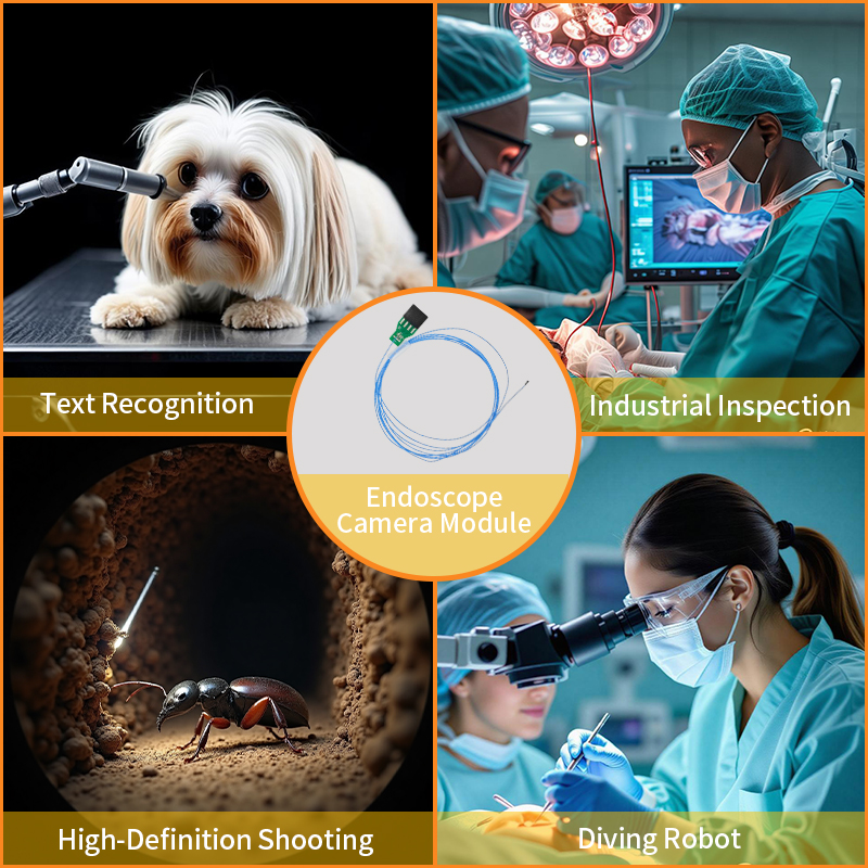
In modern minimally invasive medicine, endoscopes are key tools for doctors to observe internal human lesions and conduct precise diagnosis and treatment. The endoscope camera module, like the "visual nerve" of an endoscope, directly determines the clarity, stability, and applicable scenarios of medical images. The high-performance endoscope camera module under SincereFull, with its ultra-miniature size, high-quality imaging performance, and strong compatibility, has become an ideal adaptive component for medical endoscopes in multiple fields such as gastroenterology, respiratory medicine, urology, and otolaryngology. Its core parameters—0.9mm lens diameter, 400x400 resolution, 120° field of view, 30FPS frame rate, etc.—provide precise support for the needs of different types of endoscopes.
Core Endoscope Types Adaptable to the Camera Module
1. Gastroenterology: Gastroscopes and Colonoscopes
Gastroscopes and colonoscopes in gastroenterology are "golden tools" for diagnosing diseases of the esophagus, stomach, and intestines. The characteristics of this camera module are highly consistent with the needs of such endoscopes:
·Size Adaptability: Gastroscopes need to enter the esophagus and stomach cavity through the mouth, while colonoscopes enter the intestines through the anus. Both have strict requirements on the size of the probe. With a lens diameter of only 0.9mm, the module can be easily integrated into the probes of small-diameter gastroscopes and colonoscopes, avoiding additional irritation to the digestive tract mucosa and meeting the core demand of minimally invasive diagnosis and treatment.
·Imaging Support: The interior of the digestive tract has low light, and it is necessary to observe subtle mucosal lesions (such as inflammation, ulcers, and polyps). The 1.008μm×1.008μm pixel size of the module enhances light-sensing capability, enabling it to capture clear color images even in low-light environments. The 400x400 resolution can accurately present changes in mucosal texture, helping doctors identify early lesions.
·Dynamic Observation: The digestive tract undergoes continuous peristalsis. The maximum frame rate of 30FPS of the module ensures smooth and non-lag dynamic images, allowing doctors to clearly observe changes in the shape of the mucosa during peristalsis and avoid missing key information due to image delay.
2. Respiratory Medicine: Adaptable to Bronchoscopes
Bronchoscopes are mainly used to observe lesions in the trachea, bronchi, and intrapulmonary branches, and are important equipment in respiratory medicine for diagnosing diseases such as pneumonia, lung cancer, and airway foreign bodies. The design of this module perfectly matches its application scenarios:
·Advantages of Small Diameter and Wide Field of View: The diameter of bronchial branches gradually decreases as they go deeper. The 0.9mm ultra-miniature lens of the module can go deep into more delicate airway branches along with the bronchoscope probe. At the same time, the 120° field of view expands the observation range, reduces airway blind spots, and helps doctors fully check whether the airway mucosa has congestion, edema, foreign bodies, or tumors.
·Convenience of Focusing and Transmission: During bronchoscope examinations, doctors often need to accurately observe suspicious areas (such as nodules and ulcers). The manual focusing function of the module allows flexible focus adjustment to ensure clear details of lesions. Equipped with a Type-C interface and supporting the UVC protocol, it can quickly transmit airway images to external monitors, facilitating doctors' real-time judgment of the condition. It also supports image storage, providing a basis for subsequent diagnostic analysis.
3. Urology: Adaptable to Cystoscopes and Ureteroscopes
Cystoscopes in urology are used to examine the inner wall of the bladder, and ureteroscopes are used to observe the ureters and renal pelvis. Both need to operate in the narrow urinary tract. The characteristics of this camera module make it an ideal adaptive component:
·Adaptability of Separated Design: The probe structures of different models of cystoscopes and ureteroscopes vary. The separated design of the module facilitates flexible installation and debugging according to equipment requirements, reducing the difficulty of adaptation and improving equipment compatibility.
·Size and Imaging Guarantee: The urinary tract has a narrow space. The 0.9mm lens diameter of the module can move flexibly in the bladder and ureters, avoiding damage to the urinary tract mucosa. The 120° field of view can cover a larger range of the bladder inner wall or ureteral lumen. Combined with 400x400 resolution, it can clearly identify lesions such as stones, tumors, and inflammation, providing precise visual guidance for minimally invasive lithotripsy, tumor biopsy, and other operations, and improving the safety and accuracy of surgery.
4. Otolaryngology: Adaptable to Laryngoscopes and Nasal Endoscopes
Laryngoscopes in otolaryngology are used to observe the pharynx and larynx (such as vocal cords and pharyngeal mucosa), and nasal endoscopes are used to examine the interior of the nasal cavity and paranasal sinuses. These cavities have small spaces and dim light, and the performance of the module just meets the needs of their diagnosis and treatment:
·High Light Sensitivity for Low-Light Environments: The nasal cavity and pharynx-larynx are closed cavities, and natural light is difficult to enter. The 1.008μm×1.008μm pixel size of the module has excellent light-sensing capability, enabling it to capture clear images in low-light environments and helping doctors observe lesions such as nasal mucosa congestion, pharyngeal-laryngeal inflammation, and vocal cord polyps.
·Dynamic Imaging Aids Precise Diagnosis and Treatment: During laryngoscope examinations, it is necessary to observe dynamic processes such as vocal cord vibration. The 30FPS frame rate of the module ensures smooth dynamic images, avoiding misjudgment by doctors due to image lag. At the same time, the 0.9mm lens diameter can easily enter small cavities such as the nasal cavity and pharynx-larynx. Equipped with a manual focusing function, it can accurately focus on specific areas such as vocal cords and sinus openings, providing clear visual support for minimally invasive surgeries such as vocal cord polyp resection and sinusitis treatment.
Conclusion
The reason why this endoscope camera module of SincereFull can be adapted to the core endoscopes in gastroenterology, respiratory medicine, urology, and otolaryngology lies in the high matching between its parameter design and the needs of medical scenarios: the ultra-miniature 0.9mm lens diameter meets the minimally invasive needs of small-diameter endoscopes in various fields; the high-quality imaging performance (high resolution, high light sensitivity, high frame rate) ensures the clarity and stability of images in different environments; the separated design, Type-C interface, and UVC protocol improve equipment compatibility and ease of use. Whether it is the digestive tract, airway, urinary tract, or otolaryngological cavities, this module can rely on its core advantages to provide precise and reliable "visual support" for endoscopes, helping doctors carry out more efficient and safer minimally invasive diagnosis and treatment.
If you are interested in such products or looking for a one-stop solution for endoscope camera modules, please feel free to contact us at SincereFull!


